| PMC full text: | Published online 2018 Jan 16. doi: 10.3390/ijms19010268
|
Figure 1
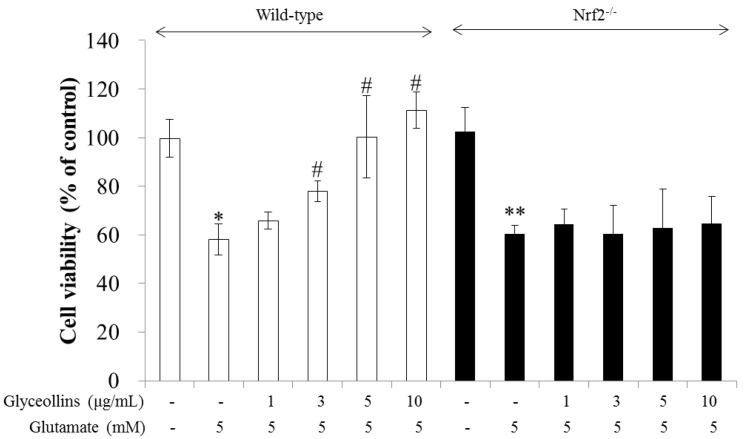
Attenuation of glutamate-induced excitotoxicity by glyceollins in primary cortical neurons. Cortical neurons isolated from fetal brain of C57BL/6 wild-type mice (white bars) or Nrf2 knockout (black bars) mice were treated with 1–10 μg/mL of glyceollins in the presence of glutamate for 24 h and followed by MTT assay. Bars represent mean ± standard deviation (SD, n = 4). *, **, Statistically significant difference from the untreated control (p < 0.05). #, Statistically significant difference from the positive control group treated with glutamate alone (p < 0.05).
