|
Anatomic Relation Between the Rectus Capitis Posterior Minor Muscle
and the Spinal Dura Mater
Thanks to Rick Hallgren for the use of these articles!
Gray's Anatomy states that the posterior atlanto-occipital membrane is in relation with the rectus capitis posterior minor (RCPMI) muscle dorsally and with the spinal dura, to which it is "intimately adherent" ventrally (See Figure 1). Nowhere in this edition is any functional relationship described between the RCPMI muscle and the dura mater. The literature seems to have missed entirely the anatomic relationship described in this present paper.

Figure 1
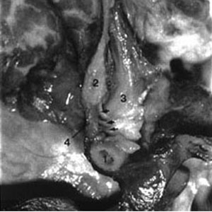
A head and neck specimen obtained from a fresh unembalmed human adult male cadaver was procured from the Maryland State Board of anatomy. A midline sagittal section was performed. Specifically studied was the RCPMI muscle and its relationship to the dura mater. The RCPMI muscle was immediately visible, arising from the posterior arch of the atlas and ascending to be inserted into the surface of the occipital bone from the inferior nuchal line to the foramen magnum (See Figure 2). A well-organized connective tissue interface was observed passing from the RCPMI muscle through the atlanto-occipital joint and inserting onto the spinal dura via the posterior atlanto-occipital (PAO) membrane.

Figure 2
We observed that the PAO membrane was securely fixed to the surface of the dural tube by multitudinous fine connective tissue fibers. There was no real interlaminar space between these two structures and they appeared to function as a single entity. The influence of the RCPMI muscle on the dura mater was artificially produced in the hemisected specimen. Artificially functioning the muscle produced obvious movement of the spinal dura between the occiput and the atlas, and resultant fluid movement was observed to the level of the pons and cerebellum. Brain tissue shrinkage occurred rapidly upon dissection and with the passage of time the observable fluid movement was not as broad, but still evident. Once the brain tissue was removed, functioning the muscle again produced observable changes in the position and tension of the dura mater. These again produced observable changes in the position and tension of the dura mater. These changes were noted within the spinal dura, as well as the dura of the posterior cranial fossa. This would account for the observed fluid movement to the level of the pons and cerebellum.
It has been suggested that the function of the RCPM muscle is to provide static and dynamic proprioceptive feedback to the CNS, monitoring movement of the head and influencing movement of the surrounding musculature. We suggest that the RCPMI muscle may also act to monitor and control movement (such as folding) and tension of the spinal dura mater. In head extension the dura folds or pleats, the greatest amount of folding would occur theoretically in the area capable of the greatest degree of extension which would be the atlanto-occipital joint. This potential dual monitoring system of the RCPMI muscles may act to prevent dural folds thus protecting cerebrospinal fluid hydrodynamics (flow) during head extension.

Return to HEADACHE
Return to ChiroZINE ARTICLES
|