
Spondylotherapy and Related Vibropercussion Therapy
© 1999. R. C. Schafer, DC, PhD, FICCINTRODUCTION
When patients enter a DC's office, they seek relief. Certainly, both patient and doctor want to know the cause of their problem (a diagnosis), but their priority is to find relief --and as rapid as possible. It is thus the attending doctor's goal to offer a rational means for the relief of pain and disability even before the diagnosis is firmed when possible. And more often than not, the patient's entering complaint will be almost resolved before the final diagnosis is determined. Of course, the relief of discomfort is just the initial stage of healing the cause of the complaint.
Gaining a reputation of offering such services is the basis of sound practice building and community relations. Media advertising cannot do it. It only announces to the public that the doctor is in need of patients. Degrading promotional gimmicks such as offering "free this" or "free that" cannot do it. It only broadcasts that the doctor's services have little value. Quickly satisfying patients' priority needs within the doctor's authorized scope of practice is the only basis for a satisfied patient recommending the practitioner responsible to a friend, relative, or colleague. It is the sole basis on which patient loyalty can be assured.
Although adjunctive procedures such as spondylotherapy are described in chiropractic case management protocols, it should always be remembered that the articular adjustment is the core of chiropractic therapy. Ancillary procedures can condition tissues to receive and respond to articular therapy and enhance physiologic mechanisms, but, with infrequent exceptions, they should not be considered substitutes.
"No instrument or machine has been invented that can replace the analytical and healing power ignited by a skilled practitioner's integrated hands, eyes, and ears." This was the belief of the great clinical diagnostician Cabot. Although it was the basis of his astute teachings to clinicians about 100 years ago, it is as true today as it was then.
It should be recognized that this author appreciates the value of numerous physiotherapeutic applications along with nutritional guidance, counseling, and standardized rehabilitative procedures. Yet they all stand in the shadow of the basis for and the proper administration of the skillful manual chiropractic adjustment.
D. D. Palmer founded chiropractic on the basis of maintaining neurologic homeostasis --a state he called "tone," as so announced to the world on the title page of his only book. Thus, any method that aids the achievement of this goal is in parallel with the basic premise of chiropractic whether it be a nonincisive surgical procedure such as an articular adjustment, spondylotherapy, heat or cold, electric stimulation, therapeutic nutrition, chemotherapy, major surgery or whatever. The manual adjustment is not the exclusive means of restoring health. It just happens to be the best means discovered so far to restore neural dysfunction at the articular level, which is so often the basis of pain and disability.
Despite what some might believe, no therapy is noninvasive and no therapy is devoid of chemical reactions. Any external stimulus applied to the body (even light touch) ignites multimillions of impulses from the periphery to the CNS where they are attenuated, dampened, correlated, synergized, integrated, and/or harmonized by neurochemical processes.
The error of allopathy is not in its use of chemotherapy, and DCs who use this argument to defend their position are in error. Our entire body is composed of chemicals. The water we drink, the food we eat, and the air we breath to survive are nothing but chemical compounds. No, the error of allopathy is not in its use of chemotherapy. It is in its use of compounds whose side effects may be more harmful than beneficial to the patient and its overemphasis of the power of common environmental microorganisms within a resistant host.
A chiropractic adjustment ignites a multitude of chemoneurologic mechanisms at the intervertebral foramen, on the spinal cord, along the neuraxes, and within the synapses and axoplasmic flow of the involved neurons. Spondylotherapy enhances this effect.
APPLYING SPONDYLOTHERAPY
Most clinical chiropractic students are introduced to the origin and basis of spondylotherapy in NCC's classic text Chiropractic Principles and Technic. The description presented therein refers to application within pioneer chiropractic. Nevertheless, it has served as a foundation for further research. We have progressed far from the block and hammer technique used in the 1920s.
The application of spondylotherapy is based on spinal cord function, especially the autonomic mechanisms, and takes the view that "the spinal cord is not a single contiguous structure but consists of 31 segments or neuromeres, each one of which serves as a little 'reflex brain' and from which point many of the vegetative (involuntary visceral) and even many somatic activities are initiated. These neuromeres serve both as the CNS part of a simple reflex arc and as a focal point for initiating remote synergistic physiologic processes often via the long tracts.
STIMULATION OF SPINAL CENTERS
To apply spondylotherapy, the clinician must be well acquainted with spinal cord function and visceral innervation. At one time, chiropractic students were required to memorize data similar to that shown in Table 1.
Deep, repetitive, short-duration percussion upon a neuromere, usually applied today by an electric vertical percussion vibrator at a rate of 1–3 impulses per second for about 20 seconds with a similar rest interval is used to stimulate a spinal center. Total session duration for excitation is usually 1–2 minutes. Pressure should be held firm to avoid slippage but not be excessive. Prolonged stimulation (over 3 minutes) tends to fatigue neuromere excitability and produces an inhibitory effect. See Tables 2 and 3.
Spondylotherapy can be applied manually by placing a clenched first over the appropriate spinal region and repeatedly striking it with the other closed fist. Decades ago, electrotherapists used a paraspinal sinusoidal current to apply spondylotherapy. In more recent years, pulsating ultrasound has been investigated.
A large number of types of visceral distress can be relieved in this manner; however, the effect is usually temporary. But not infrequently, the relief is relatively permanent. This has been attributed to breaking a self-perpetuating reflex arc.
Only two of the cephalad parasympathetics can be directly influenced by spondylotherapy: The vagus nerve at the C1-2 level and the phrenic nerve at C3-4. Vagal stimulation is usually applied to increase gastric activity, intestinal peristalsis, and nasal secretions. Phrenic inhibition is helpful in chronic cough and hiccups.
Table 1. Effects of Induced Sympathetic and Parasympathetic Stimulation (eg, Spondylotherapy
Sympathetic Division Parasympathetic Division
Structure Supply
Effect of Stimulation Supply
Effect of Stimulation Thyroid gland T1
Increases secretion X
Decreases secretion Parathyroids T1
Increases secretion X
Decreases secretion Mucous mem- branes of the head T1–2
Vasoconstriction VII
Vasodilation Salivary glands T1–2
Increases organic substances IX
Increases watery substances Pupils T1–2
Dilation III
Constriction Lacrimal glands T1–3
Vasoconstriction VII
Secretion Heart T1–5
Increases rate and force of contraction, dilates coronary arteries X
Decreases rate and force of contraction, contracts coronary arteries Upper limbs T1–6
Vasoconstriction, sweating, piloerection ? (unknown) Bronchi and lungs T1– 7
Dilation, vasoconstriction X
Constriction, vasodilation Sphincter of Oddi T4–8
Constricts X
Relaxes Gallbladder T4–8
Relaxes muscle, constricts sphincter X
Constricts muscle, relaxes sphincter Stomach T5–9
Decreases secretion and motility X
Increases secretion and motility Spleen T6–8
Contracts smooth muscle X
Relaxes smooth muscle Pancreas T6–9
Decreases secretion X
Increases secretion Liver T8–10
Increases glycogen to glucose, protein metabolism; vasoconstriction X
Opposite Pyloric sphincter T9
Increased tone, contraction X
Relaxation Adrenals T9–10
Increases secretion X
? (unknown) Small intestine T9–L1
Slightly decreases peristalsis and secretions; vasoconstriction X
Increases peristalsis and secretions, relaxes sphincters Kidneys T10–L1
Vasoconstriction, inhibits X
? (unknown) Prostate T10–L1
Contracts muscle and spermatic vein S2–4
Increases secretion Fallopian tubes T10–L1
Contracts muscle ? (unknown) Urinary bladder T12–L2
Constricts sphincter, relaxes wall S2–4
Relaxes sphincter, constricts wall Lower limbs T12–L2
Vasoconstriction, sweating, piloerection ? (unknown) Uterus L1
Contracts body S2–4
Relaxes body, contracts cervix Ileocecal valve L1
Contracts S2–4
Relaxes Penis, clitoris L1–2
Duct contraction, ejaculation S2–4
Erection Colon and rectum L1–3
Decreased peristalsis S3–5
Increased peristalsis Anal sphincter L3
Contracts S3–5
Relaxes
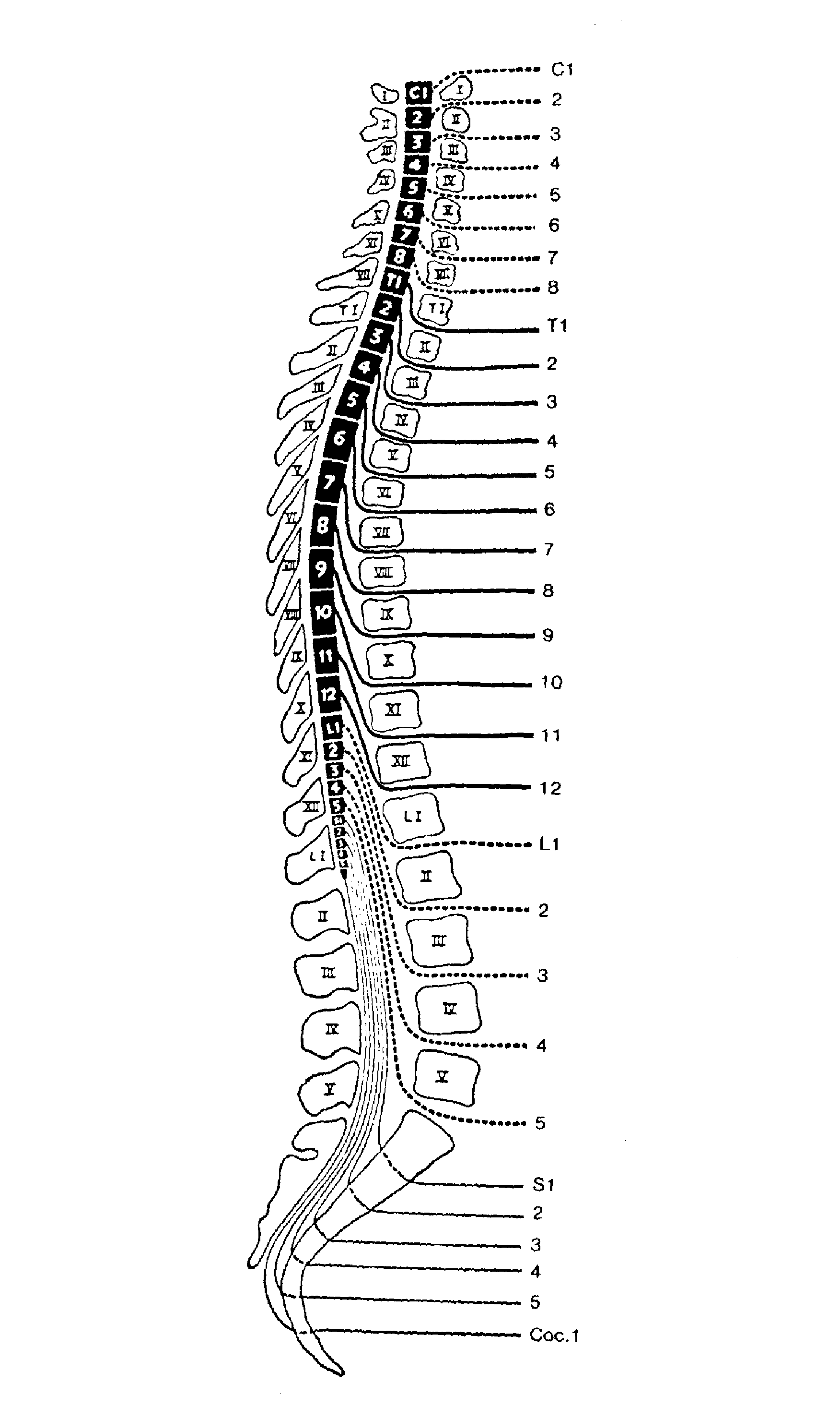
NOTE: Supply segments may vary slightly depending on the authority used. The above is typical of most studies researched. Individuals' anatomy often refuse to follow textbook models. Ask any surgeon. It is also well to keep in mind that a midthoracic spinous process, say T7, is about one level below the T7 neuromere. All five sacral neuromeres are located at the T12–L1 levels. See Figure 1.
Table 2. Duration Guidelines for Vibropercussion Therapy
Concern Time (min)
Comments Spinal center stimulation 1-2
Use low velocity with firm pressure. Monitor patient at 1-min intervals. Spinal center inhibition 4-5
Use sustained high velocity. Monitor patient at 2-min intervals.
Miscellaneous Applications
Concern Time (min)
Comments Localized treatments 10 or less
Use more time for chronic conditions, less time for acute and subacute disorders. Reducing trigger points 6–8
Too long or too strong treatment can retraumatize the site and possibly bruise the surrounding area. Relaxing muscles 2–10
Use high-velocity low-force setting. Postural drainage 3–15
Duration varies considerably depending on the particular condition being treated. General body relaxation 3–5
Use mild high-velocity force over the whole spine. Moderate toning massage 10–12
Duration varies considerably, depending on the particular condition being treated.
Table 3. Variable Effects of Vibropercussion Speed in Miscellaneous Applications
Velocity Effects High Analgesia and anesthesia by production of dynorphins and enkephalins.
Decrease trigger points
Muscle and periarticular tissue goading
Spasticity relaxation
Pre-exercise or stretching conditioning
Superficial circulatory stimulationMedium Similar effects as high but used when a milder effect is desired. Low Decongestion
Edema reduction
Anesthesia (mild)
Myotonia
Venous and lymph drainage enhancement
Relationship of Vertebra Level and Neuromere Level
In the embryo, the spinal cord extends the entire length of the vertebral canal. Nerve roots then extend laterally from the cord throughout. However, because the vertebral column grows faster than the spinal cord, the relationship of neuromere level and vertebra level does not exist during progressive growth. In the newborn, the spinal cord ends near the L2–L3 level. In the adult, it usually terminates with the coccygeal neuromere near the lower border of L1.
The length and obliquity of nerve roots increase progressively as the caudad end of the vertebral is approached because of the increasing distance between the spinal cord segments (neuromeres) and the corresponding vertebrae number. See Figure 1.
Figure 1. Schematic relationship of neuromere and vertebra level.

Return to R. C. SCHAFER MONOGRAPHS


| Home Page | Visit Our Sponsors | Become a Sponsor |
Please read our DISCLAIMER |
